


Interdisciplinary research at the nanometer scale:
AFM + Confocal Raman + SNOM + TERS
General information
Integration of SPM and confocal microscopy/Raman scattering spectroscopy. Owing to Tip Enhances Raman Scattering it allows carrying out spectroscopy/microscopy with up to 10 nm resolution.
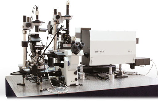
NTEGRA Spectra - AFM / CONFOCAL RAMAN & FLUORESCENCE / SNOM / TERS
Integration: The key to the new sciences
Change happens at interfaces and today’s most exciting changes in microscopy are happening where multiple technologies are interfaced together. NTEGRA Spectra is a prime example, uniting the full power of atomic force microscopy (AFM), confocal Raman and fluorescence microscopy and scanning near-field optical microscopy (SNOM) in one platform.
Simultaneous AFM and confocal Raman / Fluorescence imaging
NTEGRA Spectra supports most of the existing AFM modes (more than 30) providing comprehensive information about physical properties of the sample with nanometer scale resolution: local stiffness, elasticity, conductivity, capacitance, magnetization, surface potential and work function, friction, piezoresponse etc. Simultaneously with AFM, confocal Fluorescence and Raman measurements provide information about sample chemical composition, crystal structure and its orientation, presence of impurities and defects, macromolecular conformation, and so on.
Measurements can be performed either through upright or inverted light excitation geometries. The sample can be in a controlled atmosphere or in a liquid environment, all under controlled temperature. Complete Raman /fluorescence spectrum is recorded in each point of 2D / 3D scan with further powerful software analysis. Due to the excellent microscopy performance of the NTEGRA Spectra, 3D spectral distribution can be studied with the spatial resolution reaching the theoretical limit.
Microscopy and spectroscopy at the molecular scale
Diffraction limited spatial resolution and weakness of Raman signal are the two major challenges in Raman microscopy. When using visible light, resolution of classical confocal microscopy does not go below 200 nm. The Raman signal is often only 1/millionth of the strength of a fluorescence signal. The new world of nanotechnology has disclosed a fascinating phenomenon: the electromagnetic field can be strongly enhanced near nanometer-scale metal asperities (“nano-antennas”).
The resulting effects are called Surface Enhanced Raman Scattering (SERS) and, when done in conjunction with an SPM tip, one can get Tip-Enhanced Raman Scattering (TERS).
By using a specially prepared sharp needle tip, NTEGRA Spectra can multiply the Raman signal strength by a few orders of magnitude from a precisely scanned, localized spot on the surface several nanometers in diameter. Even single molecules can be detected and recognized by their spectra. Lateral resolution of Raman (TERS) and fluorescence maps is no longer limited by light diffraction and can be less than 15 nm.
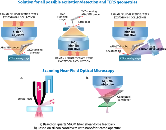
Different configuration of AFM with confocal Raman/Fluorescence microscope
|
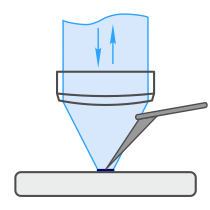
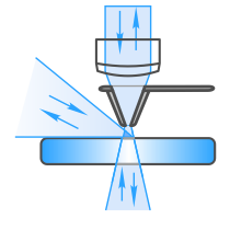
Upright A unique configuration for simultaneous AFM - Raman - TERS* and SNOM imaging of opaque samples *TERS: Tip Enhanced Raman Scattering, Tip Enhanced Fluorescence etc. |
 |
|
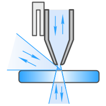
Inverted Optimized for simultaneous AFM - Raman - TERS* and SNOM imaging of samples on transparent substrates (living cells, nanoparticles etc.) |
 |
|
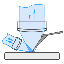
Side illumination option Used to facilitate TERS* measurements on opaque samples |
 |
|
Fiber Scanning Near-field Optical Microscopy (SNOM) SNOM techniques based on on quartz fiber. |
 |
|
Cantilever Scanning Near-field Optical Microscopy (SNOM) SNOM techniques based on cantilevers with aperture. |
 |
• Atomic Force Microscopy ( > 30 modes )
• Confocal Raman / Fluorescence / Rayleigh Microscopy
• Scanning Near-Field Optical Microscopy ( SNOM / NSOM )
• Optimized for Tip Enhanced Raman and Fluorescence (TERS, TEFS, TERFS) and scattering SNOM (s-SNOM)
New era of integration
Optical AFM (NT-MDT) + Raman spectrometer (Renishaw) = Join the best technologies in one system
HybriD Mode™
Ntegra Spectra equipped with new electronics and software allows to combine a recently developed innovative HybriD Mode™ (HD-AFM™ Mode) for nanomechanical proprieties and Raman for chemical imaging of exactly the same area within single measurement session.
 |
 |
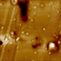 |
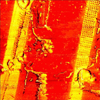
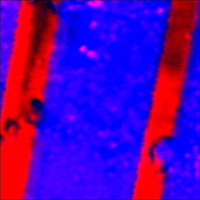
| Stiffness of HDPE/LDPE polymer sandwich cut by microtome |
Overlap of Raman maps: HDPE (red), LDPE (blue) |
AFM topography |
Working principle
Modes:
- AFM (mechanical, electrical, magnetic properties, nanomanipulation etc.)
- White Light Microscopy and Confocal Laser (Rayleigh) Imaging
- Confocal Raman Imaging and Spectroscopy
- Confocal Fluorescence Imaging and Spectroscopy
- Scanning Near-Field Optical Microscopy (SNOM)
- Tip Enhanced Raman and Fluorescence Microscopy (TERS, TEFS, TERFS)
Controlled environment:
- Temperature
- Humidity
- Gases
- Liquid
- Electrochemical environment
- External magnetic field
Applications
- Graphene, carbon nanotubes and other carbon materials
- Semiconductor devices
- Nanotubes, nanowires, quantum dots and other nanoscale materials
- Polymers
- Optical device characterization: semiconductor lasers, optical fibers, waveguides, plasmonic devices
- Investigation of cellular tissue, DNA, viruses and other biological objects
- Chemical reaction control
| Graphene flakes | Ni foil | PC-PVAC film | MoO3 |
 |
 |
 |
 |
| 30x30 um | 20x20 um | 30x30 um | 30x30 um |
Specifications
| Confocal Raman/Fluorescence microscopy |
| AFM/STM: Integration with spectroscopy |
| Software |
| Spectroscopy |
| Scanning Near Field Optical Microscopy (SNOM) |
| Optimized for Tip Enhanced Raman Scattering (TERS) and other tip-related optical techniques (S-SNOM, SNIM, TEFS, STM-LE etc.) |
| Confocal Raman/Fluorescence microscopy |
| Confocal Raman/Fluorescence/Rayleigh imaging runs simultaneously with AFM (during one sample scan) |
| Diffraction limited spatial resolution: <200 nm in XY, <500 nm in Z (with immersion objective) |
| True confocality; push button from software to control the motorized confocal pinhole for optimal signal and confocality |
| Motorized variable beam expander/collimator: adjusts diameter and collimation of the laser beam individually for each laser and each objective used |
| Full 3D (XYZ) confocal imaging with powerful image analysis |
| Hyperspectral imaging (recording complete Raman spectrum in every point of 1D, 2D or 3D confocal scan) with further software analysis |
| Optical lithography (vector, raster) |
| AFM/STM: Integration with spectroscopy |
| Upright and Inverted optical AFM configurations (optimized for opaque and transparent samples correspondingly); side illumination option |
| Highest possible resolution (numerical aperture) optics is used simultaneously with AFM: 0.7 NA for Upright, 1.3–1.4 NA for Inverted |
| AFM/STM and confocal Raman/Fluorescence images are obtained simultaneously (during one scan) |
| All standard SPM imaging modes are supported (>30 modes) — combined with confocal Raman/Fluorescence |
| Low noise AFM/STM (atomic resolution) |
| Vibrations and thermal drifts originating from optical microscope body are minimized due to special design of optical AFM heads |
| Focus track feature: sample always stays in focus due to AFM Z-feedback; high quality confocal images of very rough or inclined samples can be obtained |
| Software |
| Seamless integration of AFM and Raman; all AFM/ Raman/SNOM experiment and further data analysis is performed in one and the same software |
| Powerful analysis of 1D, 2D and 3D hyperspectral images |
| Powerful export to other software (Excel, MatLab, Cytospec etc.) |
| Spectroscopy* |
| Extremely high efficiency 520 mm length spectrometer with 4 motorized gratings |
| Visible, UV and IR spectral ranges available |
| Echelle grating with ultrahigh dispersion; spectral resolution: 0.007 nm (< 0.1 1/cm)** |
Up to 3 different detectors can be installed
|
| Flexible motorized polarization optics in excitation and detection channels, cross-polarized Raman measurements |
| Fully automated switch between different lasers — with a few mouse clicks |
| Scanning Near Field Optical Microscopy (SNOM) |
| Two major SNOM techniques supported: (i) based on quartz fiber probes, (ii) based on silicon cantilever probes |
| All modes supported: Transmission, Collection, Reflection |
| All SNOM signals detected: laser intensity, fluorescence intensity, spectroscopy |
|
SNOM lithography (vector, raster) |
| Optimized for Tip Enhanced Raman Scattering (TERS) and other tip-related optical techniques (S-SNOM, SNIM, TEFS, STM-LE etc.) |
| All existing TERS geometries are available: illumination / collection from bottom, from top or from side |
| Different SPM techniques and TERS probes can be used: STM, AFM cantilever, quartz tuning fork in tapping and shear force modes |
| Dual scan (for Hot Point Mapping in TERS): scan by sample AND scan by tip / by laser spot |
| Motorized polarization optics to produce optimal polarization for TERS |
AFM-Raman measurements can run in air, in controlled atmosphere or in liquid — all with variable temperature (for Inverted configuration)
Some features listed are optional — not included into basic system configuration
* NT-MDT AFM can be integrated with Renishaw inVia or with NT-MDT spectrometer. Specifications are given for the latter. Renishaw specifications can be found at www.renishaw.com/AFM-Raman
** Exact value of spectral resolution highly depends on how “resolution” is defined